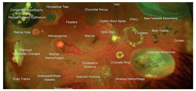
The retina and macula are the most critical parts of the eye for producing sight. These delicate structures are responsible for converting light into signals your brain can understand. When something goes wrong here, your vision—and quality of life—can change dramatically.
Common Macular and Retinal Conditions
- Age-related Macular Degeneration (AMD)
- Macular Holes
- Epiretinal Membranes (ERM)
- Retinal Detachments or Tears
- Lattice Degeneration
- Choroidal Nevus (eye moles)
- Inherited Genetic Dystrophies
Many of these conditions are silent in early stages—early detection is critical.
What’s Included in a Retinal Exam?
All the advanced imaging tools we use are designed to catch problems before vision loss begins.
Optos Ultra Widefield Retinal Imaging
This panoramic scan captures up to 200 degrees of your retina in a single image—ideal for spotting issues like retinal detachments, lattice degeneration, hemorrhages, or eye tumors. Image comparison from previous visits helps us monitor disease progression or resolution.
Retinal Photography
These high-resolution images target the macula and optic nerve—the “command center” of your vision. It’s essential for detecting early damage or structural changes over time.
Optical Coherence Tomography (OCT)
This 3D scan allows us to look below the surface of your retina, revealing hidden swelling, fluid leakage, or tissue separation. It’s indispensable for monitoring macular degeneration, diabetic macular edema, or hereditary retinal disorders.
Fundus Autofluorescence
This specialized filter technique reveals metabolic stress or subtle damage that can’t be seen with regular imaging. It helps highlight changes in pigment or waste buildup in the retina—critical in AMD and genetic disorders.
Pupil Dilation (If Needed)
Not always required, but highly valuable. When necessary—such as in suspected retinal tears or detachment—dilating the pupil gives us a full view of the peripheral retina beyond what even Optos can reveal.
Monitoring is Key
Most retinal or macular conditions don’t cause pain—and some don’t affect vision until it is too late. That’s why annual monitoring with digital imaging is critical. We store and compare data over time so you always know if things are improving, stable, or need intervention.
